the short answer first
Yes, in the right situation. If a cavity between two teeth is caught while it is still confined to the outer enamel layer, and it has not progressed into the dentin underneath, I can sometimes monitor it with fluoride treatment and repeat bitewing X-rays instead of drilling right away. The qualifier that matters most is depth, not size on the X-ray. Once decay reaches the dentin, enamel can no longer rebuild itself over that spot, and a filling becomes the realistic option. The only way to know which stage a specific lesion is in is an in-person exam with current bitewing X-rays, since two dark spots that look similar can be at very different stages.
why the space between two teeth decays quietly
A cavity that forms between two teeth, what dentists call an interproximal or proximal lesion, develops in a spot no toothbrush bristle and often no amount of good brushing alone can fully reach. Plaque sits against the tight contact point where two teeth touch, feeding on sugars and starches and slowly demineralizing the enamel from the side rather than from the top of the tooth. Because this happens below the contact point, it is usually invisible to the eye and to a mirror check.
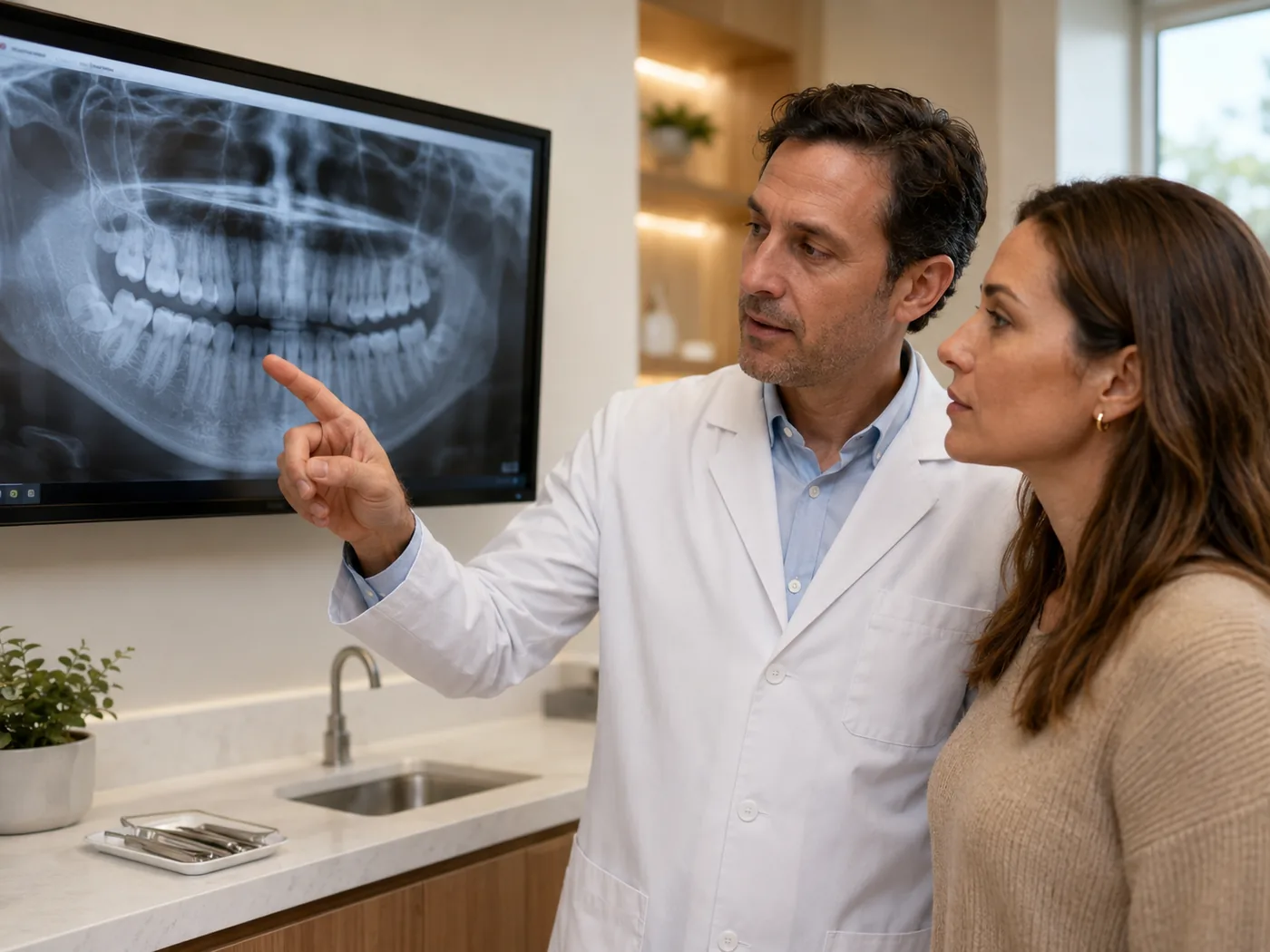
Most interproximal cavities are found on a bitewing X-ray, sometimes at a routine cleaning long before there is any sensation at all. That is part of why this topic confuses people: the first time you hear about the cavity is often the same visit where you are shown a small dark triangle on a screen, with no pain to tell you how urgent it actually is.
the assumption I hear most often about that dark spot
Many patients assume that any visible dark spot on a bitewing X-ray between two teeth automatically means a filling has to happen that day or the next. It is an understandable assumption, since most cavity conversations do end with a filling. But a very early lesion that is still confined to the outer enamel layer is different from one that has already crossed into the dentin underneath.
Enamel has no nerve supply and no living cells, yet it can still gain back minerals it lost through exposure to fluoride and saliva. That is the mechanism behind why a lesion caught at this exact stage is sometimes something we watch rather than something we drill, and why the same-looking dark spot does not always mean the same treatment plan.
what actually tells me enamel-only from already-in-the-dentin
On a bitewing X-ray, I am judging where the dark area sits relative to the boundary between enamel and dentin, not just how dark or how wide it looks. A lesion that stays within the outer half of the enamel is treated very differently from one that has already reached the dentin, even if both look similar in size at first glance.
When a lesion looks confined to enamel, I do not treat one image as the final word. I will typically monitor it over consecutive visits, comparing new bitewings against the earlier ones, watching for whether it is stable, shrinking, or advancing, and pair that monitoring with a professional fluoride treatment. Once a lesion has clearly reached the dentin, that comparison stops being useful, because remineralization is no longer a realistic option at that depth, and a filling is the appropriate next step.
what can genuinely support a spot like this
If you are told an area is being monitored rather than filled, the months between visits are not just waiting. Certain habits meaningfully support enamel's ability to rebuild itself before the next check.
- Using a fluoride toothpaste and, if recommended, a prescription-strength fluoride rinse
- Flossing that specific contact point daily, even though it cannot fully clean the spot itself
- Cutting back on frequent snacking and sipping, since enamel needs recovery time between acid exposures
- Keeping the scheduled follow-up X-ray rather than assuming no pain means no need to go back
- Asking specifically which tooth and which surface is being watched, so you know what to expect next
An early lesion that is still in the enamel is one of the few stages of a cavity that can genuinely move backward instead of forward.
when this stops being a wait-and-see situation
Monitoring only makes sense while three things stay true: the lesion stays confined to enamel on repeat imaging, there is no new sensitivity to cold, sweets, or biting pressure, and the follow-up visits are actually happening on schedule. Any new pain, a shadow that now clearly reaches the dentin on X-ray, or a lesion that has visibly grown since the last image changes the plan.
Pain from this specific area, in particular, usually means the decay has already reached the dentin or is close to the nerve, and that is not something to wait out until your next cleaning.
filling, monitoring, or something in between
When a filling is the right call, most interproximal cavities are treated with a conservative composite filling shaped to restore just the affected surface, not the whole tooth. For a lesion caught right at the enamel-dentin border, some cases qualify for a more conservative option, such as a sealant or resin infiltration, meant to arrest the lesion without a full filling, though not every case is a candidate.
When monitoring is appropriate, it is genuinely not a lesser choice. It is the option that avoids restorative work on a tooth that may not need it yet, provided the follow-up actually happens on the schedule your dentist sets.
questions patients often ask me about this
How long can a dentist monitor a cavity before it needs a filling? There is no fixed timeline. It depends on comparing consecutive X-rays, usually spaced roughly six to twelve months apart, to confirm the lesion is genuinely stable rather than assuming it.
Can brushing harder shrink a cavity between my teeth? No. Brushing more aggressively does not reach the contact point and can wear at the gumline instead. Flossing and fluoride are what actually support that specific spot.
Does a cavity between teeth always show up before it hurts? Often yes, on a bitewing X-ray, since the space between teeth is not visible to the eye and frequently causes no sensation until decay is deeper.
If one proximal cavity was monitored successfully, will future ones be too? Not necessarily. Each lesion is judged on its own depth and progression, not on what happened with a different tooth in the past.
Is resin infiltration the same as a filling? No. It is a different, more conservative procedure used only for specific early lesions, not a substitute for a filling once decay has reached the dentin.
the takeaway for a spot like this
A cavity between two teeth does not automatically mean a filling the same day it is found. What decides that is depth, specifically whether the lesion is still confined to enamel or has already reached the dentin, not how dark or how large it looks on a single image. That distinction can only be made with a current X-ray and an in-person exam, and it is worth asking directly which stage your specific lesion is in before assuming a filling is the only path forward.
Related reading
Two related topics worth a look: a lingering metallic taste after a filling and bonding versus a filling for a chipped front tooth. A couple more related reads: an old filling that suddenly feels sharp and how much natural tooth veneers actually require removing. For the broader topic list, see the full dental topics index.